Who Found That Animals Are Composed Of Cells
This page contents
i. Introduction
2. XVII century
3. XVIII century
4. Nineteen century
5. 20 century
Northowadays we agree that organisms are made up of cells, but reaching this conclusion was a long way. The size of most cells is less than the resolving ability of the human eye, which is approximately 200 micrometers (0.ii mm). The resolving power is the ability to distinguish two close points. Therefore, to observe cells it was necessary the invention of devices with higher resolving power than the human being eye. This device was the light microscope. Light microscopes use visible light and glass lenses to increase the resolution power. The maximum resolving power of low-cal microscopes is 0.ii microns, a thousand times higher than that of the human eye. Just, even with the help of light microscopes, it took a long fourth dimension to realize that cells were the units that brand upwardly all living organisms. This was due to the diversity of jail cell shapes and sizes, and besides to the poor quality of the lenses that were role of the light microscopes crafted at that time. Some other issue was the lack of histological techniques to process and report fauna and institute tissues.
ane. Introduction
The ancient Greeks proposed that affair is divided into very small units. Leucippus and Democritus wrote that affair is composed of modest parts named atoms (without parts), which tin can no longer exist divided. However, others, such as Aristotle, defended a continuity of the matter, with no empty spaces. Upwardly to the 18th century, when scientists and philosophers discussed about both living and not-living matter, they supported the theory of atoms or the theory of the continuity of matter.
The history of the discovery of the smallest units of which living organisms are made up is the history of the discovery of the cell. It began when glass lenses and the devices to concur them were first crafted; in other words, it began with the invention of light microscopes at the beginning of the 17th century. From the fourth dimension of the kickoff microscopic observations to the present mean solar day, the concept of cell has evolved under the potent influence of the development of microscopes, therefore with the improvement of engineering science. It is curious, however, that the purpose of the first lenses and microscopes was to verify the quality of fabrics, not to study living organisms.
The following are some of the most important advances and concepts in the history of the discovery of the cell:
2. XVII century
1590-1600. A. H. Lippershey, Z. Janssen and H. Janssen (father and son). They are credited with the invention of the first compound microscope. 2 magnifying lenses were placed on each side of a tube. The development of this device and the lens quality improvement would later allow the visualization of cells.

1610. Galileo Galilei described the insect cuticle. He transformed a telescope into a microscope by changing the positions of the lenses. And so, he independently invented the compound microscope (he did not know well-nigh Janssen's piece of work). 1625. Francesco Stelluti described the surface of the bee body. By that time, but surfaces were observed, just non tissue sections.
1644. J. B. Odierna studied and described the first dissections of animals.
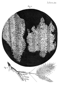
1664. Robert Hooke (physicist, meteorologist, biologist, engineer, and architect) published a book entitled Micrographia, which describes the beginning evidence for the existence of cells. He studied the cork and observed an arrangement of plant tissue resembling a honeycomb. Each piddling compartment was given the proper name of cell, only he was not enlightened that this cell (from the Latin "cella": small room) was something like what we know today as a cell. Actually, he believed that these cavities were places where found nutrients were transported. Although, he did not realize that those cells were the functional units of living things, the give-and-take cell was kept to name the petty chambers present in every plant, and then it was used to name the piffling units too found in animals.
1670-1680. North. Grew and M. Malpighi studied many species of plants and described a similar microscopic organization for all of them. In the same style every bit R. Hooke, N. Grew described institute tissues, simply the piffling chambers were regarded every bit fermentation bubbling (as in bread). He introduced the name parenchyma for some plant tissues and fabricated many drawings near the system of plant tissues. M. Malpighi gave names to many found structures such every bit trachea (due to its similarity to the trachea of insects). He also worked on animate being tissues and studied the capillary network, merely these were very rudimentary studies. These authors established the detailed microscopic arrangement of plants. However, they withal did not pay much attention to the cells, which looked similar simple air chambers.
Asouth a curiosity, dissimilar Malpighi, who thought that cells were isolated spaces, Grew proposed that the cavities of the cells were like the spaces left by threads. Thus, Grew compared microscopic organization of institute tissues with that of fabrics. Information technology has been suggested that this misunderstanding led to the incorrect option of "tissue" every bit a name to ascertain cells plus extracellular matrix in organisms. A similar misunderstanding kept the proper name "cell" to ascertain the functional and anatomical unit of living organisms.

By that time, the lenses were of very poor quality, with large chromatic aberrations, and microscopists used to put a lot of imagination in their descriptions. In this way, Gaurtier d'Agoty saw fully formed children in the head of a sperm cell, the homunculus. However, there was steady progress during this period in lens crafting, which resulted in greater definition and higher resolving power of the microscope. J. Huddle (1628-1704) was a very good lenses maker, and he taught A. van Leeuwenhoek and J. Swammerdam how to make high quality lenses.
It is thought that the animal cells were first observed under the lite microscope around 1673. They were blood cells. However, it is not known wether information technology was Malpighi, Swammerdan or Leeuwenhoek.
1670. A. van Leeuwenhoek crafted uncomplicated microscopes, with a single lens, but with a perfection that allowed him to reach magnifications higher than 270 times, more than than what compound microscopes could offer at that fourth dimension. He can exist regarded every bit the founder of microbiology considering he was the first to report on descriptions of bacteria and unicellular organisms. He wrote many manufactures well-nigh his observations on biological samples, more detailed than anyone earlier. He observed drops of water, blood, semen, hairs, and many more. He thought that all animals are fabricated up of multiple units, but failed to associate these units with the cells of plants. Leeuwenhoek had described animate being cells every bit "animalcules". Even later detailed microscopic studies of fauna tissues, many years passed before animal and institute cells were considered similar structures.
3. XVIII century
1757. Von Haller said that animal tissues are made up of fibers.
1759. The first attempt to put animals and plants at the same level was made by C.F. Wolf, who said that there is a fundamental unit with globular shape in all living organisms. This unit would exist globular at first, as in animals, then hollow, and later on filled with sap, as in plants. He as well said that the growth of the body of living organisms would occur by adding new cells. However, information technology is possible that what he observed with his microscopes were generally artifacts, and nonetheless, he fabricated these remarkable statements. In his work Theoria generationis he claimed that living organisms are formed as a issue of progressive development and torso structures would appear past growth and differentiation of less developed structures. This theory was contrary to another very pop theory at that fourth dimension: the preformationist theory, which suggested that a complete organism was already present inside the gametes, i.e. a fully adult tiny body was present in each gamete that would accomplish the adult stage but by increasing the size of each part.
1792. L. Galvani discovered the electric nature of musculus contraction.
1827. Thou. Battista Amici corrected many lens aberrations, allowing much amend microscopes.
4. 19 century


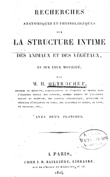
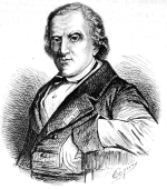
1820-1830. The development of the cell theory started in French republic with H. Milne-Edwards and F.V. Raspail. They observed a big diversity of tissues from different animals and reported that the tissues were composed of globular, but unevenly distributed units. Plants were besides regarded as having these units and also gave these vesicles a physiological part. R.J.H. Dutrochet, French also, wrote if i compares the farthermost simplicity of this hit construction, the jail cell, with the farthermost diversity of its content, information technology is articulate that it is the basic unit of an organized entity. Actually, everything is ultimately derived from the cell. He studied many animals and plants, and came to the determination that the cells of plants and the globules of animals were the same thing, just with dissimilar morphology. He also proposed some physiological functions for these units and suggested that cells are born inside other cells (non agreeing with the spontaneous generation theory). F.Five. Raspail, a French chemist, proposed that each cell is similar a laboratory that allows the organization of tissues and organisms. He thought that each cell, like Russian dolls, contained many pocket-size vesicles that could grow and exist released as independent cells. He fifty-fifty suggested that cells could have sex (more often than not hermaphrodite). Finally, he said, and not R. Virchow, "Omnis cellula e cellula", that is, every cell comes from another jail cell.
1831. R. Brown described the cell nucleus. This is rather controversial considering in a letter reported by Leeuwenhoek in 1682, he described a rounded construction inside red blood cells of fish. This structure could not be anything else only the nucleus. Yet, he did not proper name information technology. Furthermore, in 1802, F. Bauer, from the Czechia, described a cell structure very similar to the cell nucleus. Much later, M.J. Schleiden proposed that all cells contain a nucleus.
1832. B. Dumortier described binary segmentation in plant cells. He reported the synthesis of new cell wall between the two new cells and proposed that this machinery was the way cells proliferate. Thus, he refused other theories virtually jail cell proliferation such every bit those saying that cells appear inside other preexisting cells, such as Russian dolls, or those proposing spontaneous generation.
1835. R. Wagner described the nucleolus.
1837. J. Purkinje, from Czech republic, i of the best histologist at that fourth dimension, suggested not merely that animal tissues were made up of cells just also that animal tissues were analogous to found tissues.
1838. Chiliad. J. Schleiden, a German language botanist, wrote the beginning axiom of the cell theory for plants (he did non report beast tissues). That is, all plants are fabricated up of units named cells. The German physiologist T. Schwann, in his book Mikroscopische Untersuchungen, extended this concept to animals and later to all living organisms. He went beyond by saying that animals and plants follow the same bones mechanisms.
T. Schwann described cells equally units surrounded by a membrane. He actually did not find the "real" membrane of the cells. Two years before, H. Dutrochet had been suggested the presence of a membrane after his studies well-nigh osmosis. The membrane described by T Schwann, and G.J. Schleiden, actually was the cell wall plus the cortical cytoplasm of institute cells. That is why they wrongly stated that the nucleus was located within the cell "membrane". T. Schwann went farther and proposed that this "membrane" functions equally a barrier to split up the internal from the external environment, which has been proven to be correct, but for the "real" prison cell membrane.
Although the German language schoolhouse, T. Schwann and Thousand.J. Schleiden, has been recognized for the development of the cell theory statements, in that location are at to the lowest degree four researchers who wrote similar statements years earlier: Oken (1805), Dutrochet (1824), Purkinje (1834) and Valentin (1834). Dutrochet stood out amongst the other researchers. There is a rumor that T. Schwann knew about the Dutrochet's papers and "borrowed" some ideas. T. Schwann and Grand.J. Schleiden also agreed with cells emerging from the interior of other cells, but this was showon to be wrong.
1839-1843. F. J. F. Meyen, F. Dujardin and K. Barry continued and unified dissimilar branches of biological science to prove that individual protozoa are nucleated cells similar to those that make up animals and plants, and they also proposed that the continuous jail cell lineages are the basis for life. Thus, the evolutionary history of living beings could be represented in a unmarried tree of life in which plants, animals, fungi and unicellular organisms were interconnected.
1839-1846.J.E. Purkinge and H. van Mohl independently proposed the proper name protoplasm for the cell content (excluding the nucleus) of plant cells. Dujardin (1835) previously chosen it sarcode by for beast cells. It was F Cohn (1850) who realized that protoplasm and sarcode were the same affair. Studying plant and animals at same level was not common at that fourth dimension. Because plant cells showed a cell "membrane" (remember that this "membrane" was the cell wall plus cortical cytoplasm) but it could not be observed in animal cells, the living affair of cells thought to be the protoplasm. Due north. Pringsheim (1854) proposed that protoplasm is the living part of plants. At that times, virtually researchers agreed that living strength was contained in the protoplasm so that the cell membrane disappeared every bit a fundamental component of the cell. This was reasonable because the jail cell membrane can non be seen with the light microscope.
1856. R. Virchow wrote "The cell, every bit the simplest form of life-manifestation that withal fully represents the idea of life, is the organic unity, the indivisible living i". Past mid-nineteenth century this theory was widely accepted.
1879. W. Flemming described the chromosome segregation and proposed the name mitosis.
1899. C.E. Overton suggested that the interface between the protoplasm and the extracellular space was lipidic. Based on osmosis and lipid diffusion experiments, he proposed the beingness of a thin layer of lipids lining the protoplasm.
5. 20 century
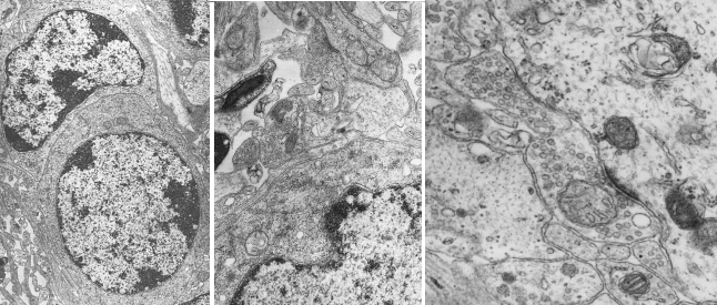
1932. The electron microscope appeared. It was invented in Germany by M. Knoll and East. Ruska, and developed during the thirties and forties of the XX century. The low-cal microscope uses visible light, just its wavelength cannot discriminate two points that are closer than 0.2 micrometers. Electron microscopy is able to written report cell structures that are as small as several nanometers (10-3 micrometers). The existence of the plasma membrane, and other inside the cell, was confirmed. It was the first time that membranes could be observed. With the help of the electron microscope, it was shown the interior of the eukaryotic prison cell is complex and rich in compartments. By 1960, the cell ultrastructure had already been studied and described.

Bibliography
Cavalier-Smith, T. 2010. Deep phylogeny, ancestral groups and the iv ages of life. Philosophical transactions of the Royal Club B. 365: 111-132
Harris, H. 2000. The birth of the cell. Yale University Press. ISBN-10: 0300082959.
Hook, R. Micrographia. 1664. Read it at US National Library of Medicine.
Ling, G. 2007. History of the membrane (pump) theory of the living cell from its beginning in mid-19th century to its disproof 45 years ago - though still taught worldwide today every bit established truth. Physiological chemical science and physics and medical NMR 39: 1–67.
http://micro.magnet.fsu.edu/alphabetize.html
Source: https://mmegias.webs.uvigo.es/02-english/5-celulas/1-descubrimiento.php
Posted by: frazieroffily.blogspot.com

0 Response to "Who Found That Animals Are Composed Of Cells"
Post a Comment